About Me
Hello! I’m Zac. I’m currently a postdoctoral scholar in the Rennison Lab at University of California - San Diego and a Lecturer at the University of San Diego. I am a broadly trained biologist with particular interest in understanding how organisms, particularly those in aquatic habitats, rapidly adapt to novel environmental and ecological conditions. Over the years I have investigated this question and related themes in a variety of study systems, ranging from tiny estuarine crabs and their barnacle parasites, a globally invasive marine tunicate, and the inimitable threespine stickleback. My research takes a holistic perspective by integrating across levels of biological organization – from molecular pathways to physiological function to ecosystem-level processes. This approach gives me the fantastic opportunity conduct fieldwork in breathtaking locations, perform molecular magic in the laboratory, and make sense of oodles of sequence data at the helm of a supercomputer. Please see my Research page if you interested in learning more.
In addition to research, my winding academic career has involved numerous teaching and mentoring experiences, culminating most recently as a part-time lecturer at University of San Diego and instructor of Summer Session coursework at UCSD. Spending time with students is an immense privilege, one that I take seriously but while bringing levity, enthusiasm, and fun to classroom. At UCSD I teach lecture-based courses, whereas at University of San Diego I lead a laboratory section. Teaching these different styles of courses, not to mention with vastly different levels of enrollment, has allowed me to try to bring the best of both methods together, using active-learning approaches in the lecture hall and fostering content mastery in the lab. Further developing my dual professional identity, of researcher and educator, is a primary focus. Please see my Teaching page if you are interested in learning more.
Below I have a bit more about my life’s path through academia if you are interested in knowing more about where I’ve been. You can also just see my CV if you’d rather skip the long story.
Academic Path
I always leaned toward the natural sciences, but it wasn’t until the the summer after my junior year of undergrad that I had the chance to get involved with research. Through an REU program at Scripps Institution of Oceanography I joined the lab of Dr. Jules Jaffe. My main focus was training an image classification software to correctly identify plankton from photos collcted by an Underwater Vision Profiler (UVP) instrument. This was combined with various tinkering projects on underwater instrumentation platforms and culminated with a five-day research cruise in the Southern California Bight. After this experience I was hooked on research and resolved to get a position in a research lab when I returned to Lewis & Clark College for my final year.
Building the skillset
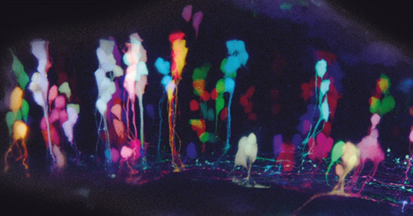
Cold emailing works, folks! I got in touch with a new professor starting in the Biology Department, Prof. Tamily Weissman-Unni, to ask about joining her lab. After our first in-person meeting upon returning to campus my fate was sealed. I’d be starting volunteering in setting up the lab, followed by a independent study project for credit, and then a full-time lab technician position upon graduation! In Tamily’s lab we studied the development of the larval zebrafish brain using fluorescence imaging techniques, namely, Brainbow. This ingenious approach that Tamily co-developed combines the expression of three fluorscent proteins with different spectral properties (i.e. colors) to create a dazzling mosaic of rainbow cells, using the same principle behind color TV (see RGB, CMYK, etc). Brainbow was pioneered as an approach in connectomics; because neighboring neurons can be easily distinguished through their unique color, fine processes like dendrites and axons could be traced. However, because we could essentially “switch on” the color blending by driving the expression the Cre recombinase with an inducible promoter, this could be carefully timed to label populations of progenitor cells in unique colors. These colors would be inherited by daughter cells and maintained through multiple mitoses, allowing for detailed mapping of cell fate. We also regularly collaborated with Prof. Vivek Unni at Oregon Health and Sciences University on developing zebrafish as a Parkinson’s disease model, focusing specifically on the protein alpha-synuclein, the main component of Lewy bodies, the pathological hallmark of the neurodegenerative disease.
In the lab I was responsible not only for the maintenance of our zebrafish colony and lab inventories, but also the fun stuff like creating new constructs for driving the Brainbow cassette in different cell-types (PCR, plasmid design, cloning, E. coli work, etc), generating transgenic lines of zebrafish (microinjections, genetic crosses, etc.), immunohistochemistry for labeling biomarkers of Parkinson’s, and, best of all, confocal imaging! Zebrafish are so cool because they are (mostly) transparent during early development, so you could actually image their genetically modified rainbow brains in vivo. Keeping a zebrafish larvae alive under the microscope for hours and hours is hard, so there was some tweaking to be done. At times we would take hours-long time lapses, being able to track individual divisions of neural progenitors, or we would return to the same spot in the same fish day after day, monitoring the expansion of cell lineages in the hindbrain.
Thankfully about half-way through my three years as a lab tech Tamily and the department were successful in securing an NSF Major Research Instrumentation award to purchase a laser-scanning confocal microscope. Part of this award was hiring a part-time Microscopy Coordinator to oversee the microscope’s use. There I was! Now I’d be spending a quarter of my time managing the microscopy suite (which included several wide-field fluorescence scopes), training users, and eventually also a sweet macrophotography system.
Although I enjoyed my time working on zebrafish neurobiology and I learned a great deal, it started to dawn on me that I would rather focus on a topic and system with more ecological and evolutionary relevance. Dissecting the inner workings of the brain was fascinating, but I was attracted to broader questions and research with more of a natural history component. I think part of this was inspired by the fact that we were just down the hall from Greta Binford’s lab at Lewis and Clark. They studied the evolution of spider toxin proteins and her trainees were jaunting off to far-flung locations to collect spiders in caves. That sounded like a lot of fun! What I really wanted to do was take all of the molecular skills I had learned and apply them to an evolutionary question.
Applying molecular tools to nature
For whatever reason, it was during this time as a lab tech that I got really set on this idea of studying parasites. I think this was at least partly inspired by the Planet Earth scene feature Cordyceps. If you haven’t seen this, look it up! Essentially a parasitic fungus infects unsuspecting ants and tweaks with their brains to compel them to climb up the tree and bite on a twig. Once they die, the fruit-body sprouts out of the ant’s head and rains it’s spores down on legions of the ant’s unsuspecting compatriots. This whole idea of parasitic behavioral manipulation just fascinated me. I started reading the literature and came across the work of Robert Poulin, among others. Again, cold emailing works! After exchanging some emails and a Skype call (no Zoom back then…), we agreed we would try to find a way for me to join him for a masters degree. Ultimately I was able to start my masters with him at University of Otago in Dunedin, New Zealand and secured funding through a fellowship specifically set aside for international students taking on research-based masters programs.
Before starting we had lots of conversations about what to work on. I had initially planned to perform experiments to get at the root of the mechanims behind behavioral manipulation. Given that I only had one year down there and that such experiments would likely take much longer, he floated the idea of doing a phylogeographic on a parasite species of my choosing. To be honest, I had no idea what phylogeography was at that point. I hadn’t even taken an evolution class in undergrad! I read and read and read, picking up classics by John Avise and others. At that point, to me molecular tools like PCR, cloning, etc. were just part of the journey towards trying to get particular zebrafish brain cells to light up. That you could use molecular data to try to resolve the history and geographic distribution of species was tremendously exciting and novel.
Ultimately during my masters research I ended up investigating the comparative phylogeography of two host-parasite systems: a mermithid nematode and its sandhopper host, and horsehair worms and their orthopteran hosts. Starting late 2015, I drove all around the southern end of New Zealand’s South Island looking for the sandhopper Bellorchestia quoyana. Grazer of decaying kelp (yum!), it could be found in the hundreds of thousands along the wrack line of a single beach. So off I went, flipping over piles of kelp and catching as many sandhoppers as I could and chucking them in a tupperware. Back at the lab, one by one I would dissect thousands of sandhoppers and look for nematodes inside. When I’d find one, I’d take some basic measurements, but then, most importantly, preserve the worm in ethanol for DNA work downstream.
At the same time, I was performing collections of horsehair worms. Yes, those. These, like the mermithids, are also expert manipulators of their hosts’ behavior. In their case, the horsehair worm drives their host to seek out water and jump in, which is usually fatal. They horsehair worm then emerges from the hosts posterior and wiggles around until it finds a mate. Notably, these worms can be extraordinarily long: we’re talking feet in just a small grasshopper (see picture of Brenah holding this bad-boy). So for this side of my masters research, rather than collecting the host and doing dissections (which is complicated because prevalence is pretty low and permitting can be a pain for some host species) I instead just collected them as free-living worms in tiny streams in the mountains of Central Otago. For whatever reason, horsehair worms are extremely abundant in New Zealand relative to other parts of the world (and much better studied). So this made my life easy. Back in the lab, same deal. Just stick them in ethanol.
CONTENT COMING
Outside of Work
CONTENT COMING